Diagnostics at L.E.C Faisalabad Branch
At Cosmetic Eye Surgeon, we believe in providing precise, advanced diagnostics to ensure the best treatment for each patient. Our array of sophisticated diagnostic tools helps in early detection, accurate diagnosis, and effective management of various eye conditions. Here’s a detailed overview of our key diagnostic services
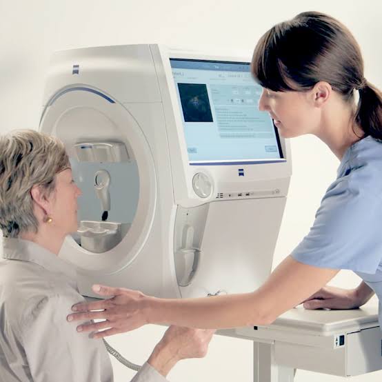
Perimetry
- Purpose: This test assesses your visual field (the total area in which objects can be seen while focusing on a central point). It is particularly useful for detecting vision loss caused by glaucoma and other optic nerve diseases.
- How it works: You will look straight ahead at a central point while lights flash in different areas of your peripheral vision. The machine measures your ability to detect these lights, helping to map areas where vision may be reduced or missing.
SD-OCT (Spectral Domain Optical Coherence Tomography)
- Purpose: SD-OCT is a high-resolution imaging technology that captures detailed, cross-sectional images of the retina’s layers. This is essential for diagnosing conditions like macular degeneration, diabetic retinopathy, and glaucoma.
- How it works: This non-invasive test uses light waves to scan the retina, providing a 3D map of the eye’s internal structures, helping doctors visualize any damage or abnormalities in the retina or optic nerve.
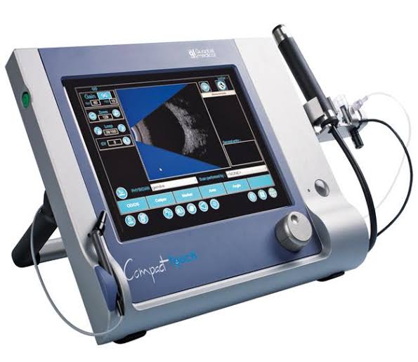
B-Scan Ultrasonography
- Purpose: B-Scan ultrasound is employed when direct visualization of the inside of the eye is not possible due to cataracts, vitreous hemorrhage, or other obstructions. It helps evaluate the retina, optic nerve, and other internal eye structures.
- How it works: A small probe is placed gently on the eyelid after applying gel, and sound waves create an image of the inside of the eye. This test is often used to assess tumors, retinal detachment, or bleeding.
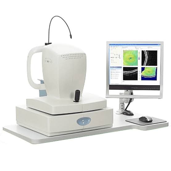
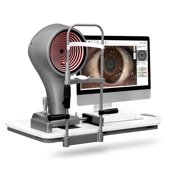
Sirius Scan
- Purpose: This diagnostic tool provides a comprehensive analysis of the anterior segment of the eye, including the cornea, iris, and lens. It’s particularly useful for detecting corneal diseases, dry eye, and for planning refractive surgery.
- How it works: The Sirius scan combines placido disc topography and Scheimpflug tomography to create a 3D map of the front part of the eye. It measures curvature, elevation, and thickness, ensuring precise assessment before surgery.
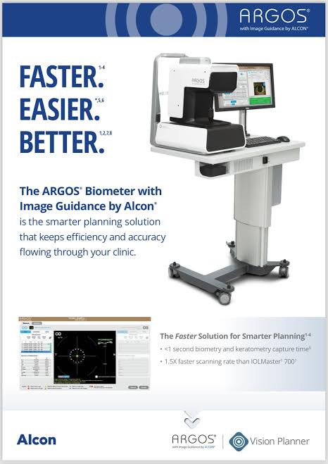
ARGOS IOL Master
- Purpose: Used primarily in cataract surgery planning, the ARGOS IOL Master precisely measures the axial length of the eye and the curvature of the cornea. This information is crucial for selecting the right intraocular lens (IOL) power to implant during surgery.
- How it works: It is a quick, non-contact method where light is used to measure the dimensions of the eye. This ensures the most accurate IOL selection, resulting in better post-surgery vision outcomes.
GALILEO Scan
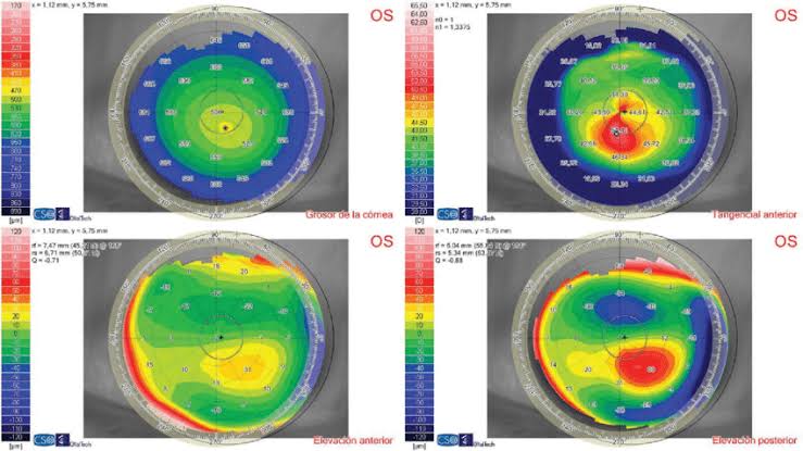
- Purpose: The GALILEO Scan provides advanced corneal mapping and pachymetry, which are vital for diagnosing and managing corneal disorders like keratoconus and for determining suitability for laser vision correction surgery.
- How it works: This technology combines various optical measurements to give a complete picture of the cornea’s shape and thickness, allowing precise planning for surgeries like LASIK or cataract surgery.
